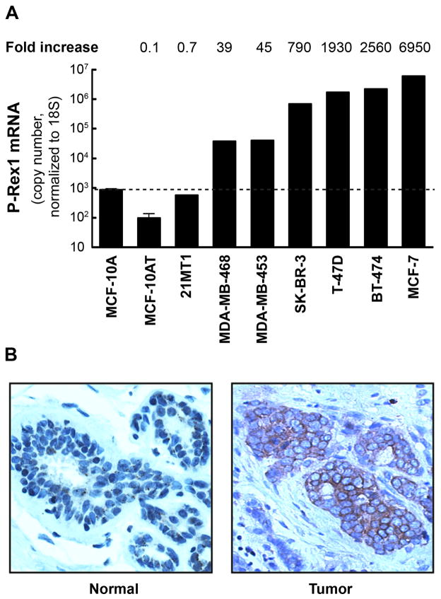
Figure 3. Overexpression of P-Rex1 in breast cancer.
Panel A. Expression of P-Rex1 mRNA by Q-PCR in human mammary cell lines. Values presented as “fold-increase” are relative to levels in MCF-10A cells. Panel B. Immunohistochemistry comparing P-Rex1 staining in a human breast tumor and normal mammary tissue.

