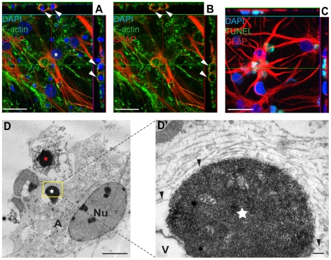
Figure 2. Astrocytes engulf dead cells following neural injury.
(A–B) Embryonic astrocytes have engulfed dead cells with highly condensed nuclei. Twenty-two hours after scratch injury, the cultures were fixed and astrocytes were identified with specific antibodies against GFAP. Phalloidin and DAPI labeling was used in order to visualize the actin cytoskeleton and cell nuclei, respectively. Condensed DAPI positive nuclei are present in cytoplasmic vacuoles (white arrows) within the viable astrocyte (white asterisk) and not in direct contact with the cytoskeleton of the astrocyte, indicating a macropinocytotic engulfment mechanism. (C) To prove that the cells with condensed nuclei found within astrocytes are de facto dead, we labeled cultures with the apoptotic marker, TUNEL. (D) TEM image of an astrocyte that appear to be in the process of engulfing a dead cell (red star). The dead cell displays the hallmark chromatin traits of apoptosis, but also secondary necrosis as seen by the ruptured cell membrane. The astrocyte contains a previously ingested dead cell (white star) in a spacious vesicle. The astrocyte is marked by an A and the nucleus is denoted Nu. (D') The area marked by a yellow rectangle in D at higher magnification. The ingested dead cell (white star) is contained in a membrane-enclosed compartment (black arrowheads) that is closely linked to the membrane on one side of the dead cell and then diverge to create the vesicle (denoted V) seen in D. Scale bars: 20 µm (A–C), 5 µm (D), 200 nm (D').

