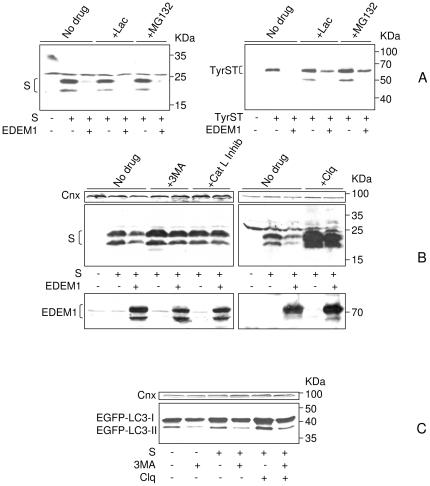
Figure 6. HBV envelope proteins are disposed by autophagy/lysosomal degradation.
HEK293T were transfected with pCiS, pTriexTyrST expressing the ERAD substrate soluble tyrosinase (TyrST), or pEGFPC1-LC3 expressing the autophagy marker LC3, in the presence or absence of pCMVEDEM1. The cells were treated with proteasomal (A) or autophagy/lysosome (B, C) inhibitors. Untreated (no drug) cells were also used as control. S, TyrST and EDEM1 synthesis was monitored by Western blotting using the corresponding Abs (A, B). Conversion of EGFP-LC3-I to EGFP-LC3-II was determined by Western blotting using anti-LC3 Abs, in the presence or absence of autophagy inhibitors (C). Calnexin (Cnx) expression was used as total protein-gel loading control.

