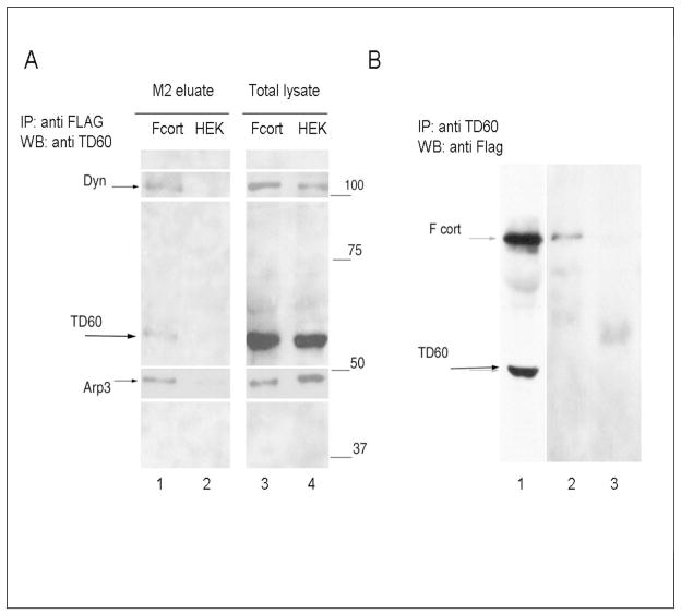
Figure 2.
Detection of TD60 by western blot following immunoprecipitation F-cortactin expressing HEK 293 cell lysates. A) Eluant of M2 agarose beads incubated with either F-cortactin expressing cell lysate (Fcort) or mock transfected HEK 293 cell lysate (HEK). Lanes 1 to 4 show the staining obtained by 2 sequential incubations of the blot beginning with anti-RCC2/TD60, followed by a mixture of mouse monoclonal antibodies specific for dynamin and Arp3. Time of exposure for each antibody was adjusted to produce the composed image shown here. B) Detection of A clarified lysate from approximately 2 × 10⁷ F-cortactin expressing cells was divided in two and treated with either a 1/200 final dilution of anti RCC2/TD60 rabbit antibody (track 2) or control rabbit serum (track 3) immunoprecipitated and analyzed in PAGE SDS and blotted with anti Flag antibody as described in Materials and Methods.

