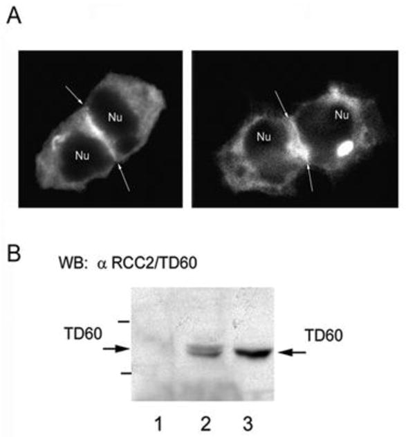
Figure 3.

A) Distribution of EGFP cortactin in diving HEK 293 cells. Two examples of EGFP-cortactin expressing HEK 293 cells 6 h after release from nocodazole driven cell cycle arrest. Arrows point to zones of marked intensity of cortactin statining in the equatorial zone of the dividing pairs of cells. B) Co-immmunoprecipitation of endogenous cortactin and RCC2/TD60 from lysates of nocodazole synchronized Hela cells. Endogenous cortactin was immunoprecipitated with monocloncal antibody 4F11 followed by western blot analysis with anti- RCC2/TD60 polyclonal antibody as described in the text. Lanes 1 and 2 corresponds to a control non-related monoclonal antibody and anti-cortactin antibody 4F11, respectively. In Lane 3 is seen an aliquot of the total cell lysate.
