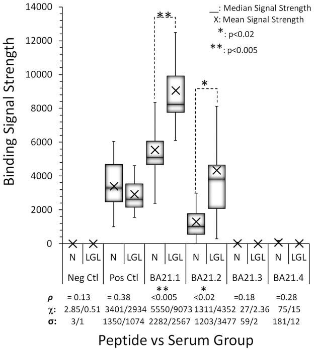
Fig 1.
Peptide Array Epitope Mapping. Overlapping peptides were tested for seroreactivity against LGL leukemia and normal donor sera (Peptide vs. Serum Group). The signal strength of the sera was interpreted as reactivity. Neg Ctl: duck hepatitis B virus core peptide. Pos Ctl: Anti-IgG1. ρ: probability, χ: mean value, σ: standard deviation.

