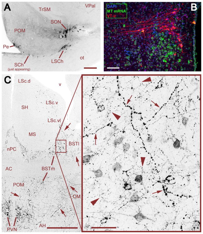
Fig. 1.
Distribution of VT-ir cell groups in estrildid finches. (A) The rostral-most cell groups in a female Angolan blue waxbill, showing VT-ir neurons in the periventricular POA (Pe), SON, suprachiasmatic nucleus (SCh), and lateral SCh (LSCh). This photo was taken rostral to the main body of cells in the SCh, thus just a few neurons are visible. Scale bar = 200 μm. (B) The PVN of a male zebra finch, showing the separation of VT and MT populations. Note that low levels of MT mRNA extend in the surrounding hypothalamus and are not restricted to the PVN. Scale bar = 50 μm. (C) VT-ir cells and fibers at the level of the anterior commissure (AC) in a male zebra finch, showing cell groups of the PVN and BSTm, and apparent overlapping projections from these cell groups in the BSTm and ventral LS. Large-caliber, heavily beaded axons (small arrows) are observed coursing from the PVN through the BSTm and directly into the ventrolateral zone of the caudal LS (LSc.vl). Relatively heavier projections are observed to the lateral BST (BSTl), but terminate immediately adjacent to the BSTm and LSc.vl. Within the BSTm (box), fine-caliber, beaded axons of local origin (large arrowheads) mix with the heavier axons of apparent PVN origin. Scale bars = 200 μm (left) and 20 μm (right). Other abbreviations: AC, anterior commissure; AH, anterior hypothalamus; LSc.d, dorsal zone of the LSc; LSc.v, ventral zone of the LSc; MS, medial septum; nPC, nucleus of the pallial commissure; OM, occipital-mesencephalic tract; POM, medial preoptic nucleus; SH, septohippocampal septum. Panel C modified from Goodson and Kabelik (2009).

