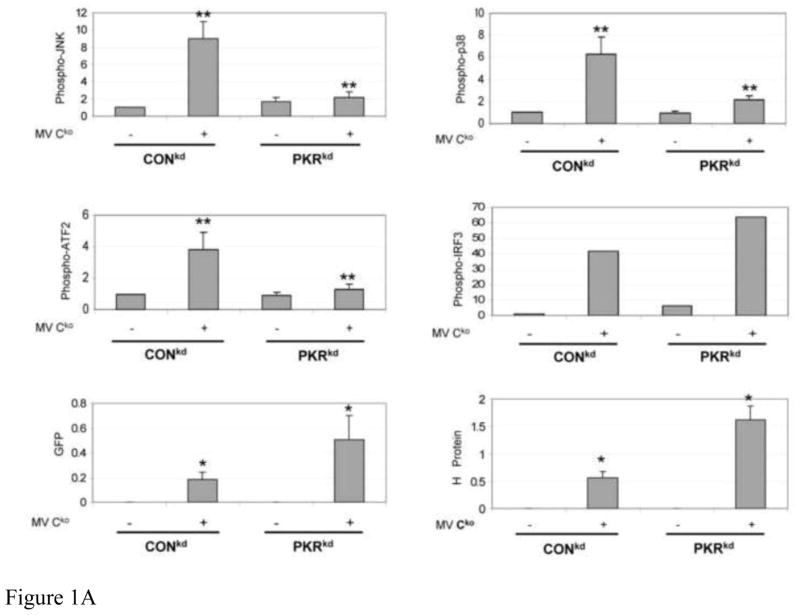
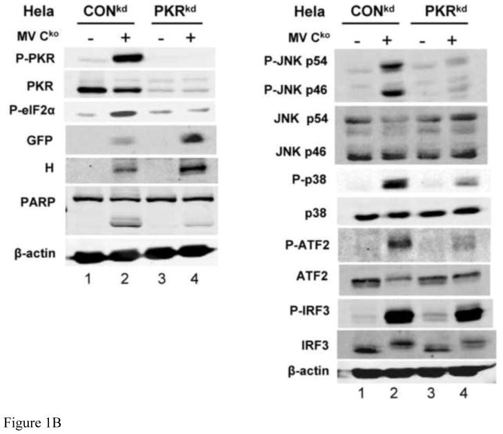
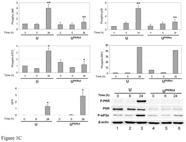
FIGURE 1. Activation of JNK, p38 and ATF2 phosphorylation is PKR dependent, but IRF3 phosphorylation is PKR independent, in Cko-infected cells.
(A) Whole-cell extracts were prepared from either uninfected (−) or infected (+) HeLa cells (CONkd or PKRkd) at 24 h after infection with Cko measles virus. Western blot analyses were performed and the blots quantified for levels of phosphorylation of JNK, p38, ATF2 and IRF3, and GFP and H protein expression, by infrared imaging as described under Materials and Methods. (B) Representative blots are also shown for PKR, phospho-PKR (P-PKR), and phospho-eIF2α (P-eIF2α), in addition to the proteins quantified in (A) above. (C) Quantitation of MAP kinase, ATF2 and IRF3 phosphorylation and GFP expression determined by immunoblot analyses of extracts prepared from amnion U cells (U) or a stable PKR knockdown clone (UPKRkd) infected for 6 or 24 h with Cko virus (6, 24), or left uninfected (0). Quantifications were performed as for Hela cells shown in (A). Representative western immunoblots for PKR, phospho-PKR (P-PKR), and phospho-eIF2α (P-eIF2α) are shown for extracts from amnion U cells, uninfected (0) or infected (6, 24 h) with Cko virus. The results shown in (A) and (C) are the means with standard deviation (n= 3). *, P< 0.05; * *, P< 0.01.


