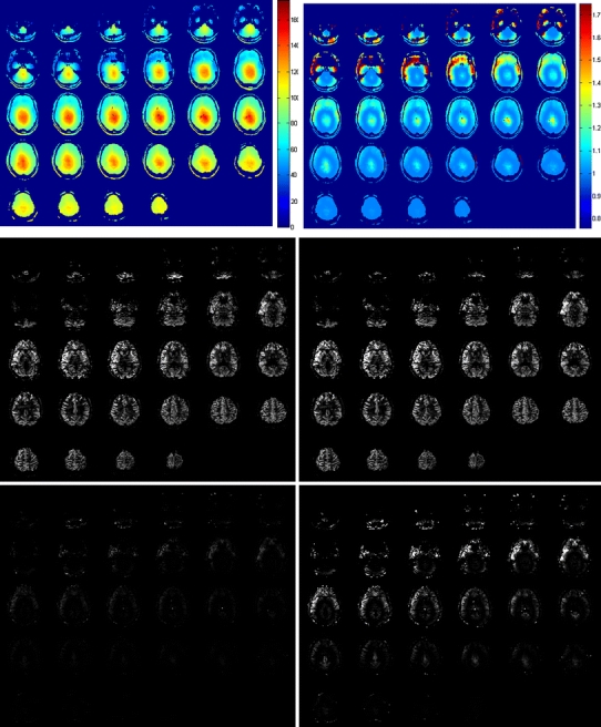
Fig. 5.

In vivo measurements of B1 efficiency and whole-brain pseudocontinuous arterial spin labeling (pCASL) measured in a female volunteer of 26 years. Top row: B1 efficiency (%) left and resulting correction factor for arterial spin labeling (ASL) images (calculated as 1/sin(90 × B1 efficiency/100%); middle row: ASL image without (left) and with (right) correction for the inhomogeneous B1; bottom row: difference of ASL images with and without B1 correction at the same contrast as the original images (left) and displayed at a five-times higher contrast level (right image)
