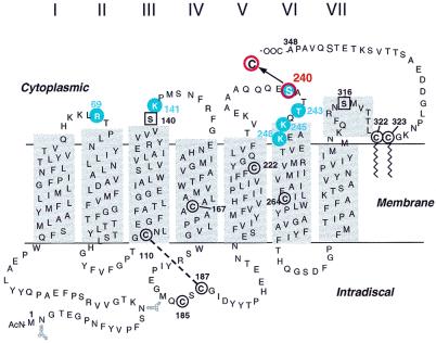
Figure 1.
A secondary structure model of rhodopsin showing positions of the single cysteine-containing mutants that were tested for attachment of R (I) and showed photocrosslinking with heterotrimeric Tαβγ (circled in blue). Mutant S240C (circled in red) was taken through all steps in the strategy described in Fig. 3.

