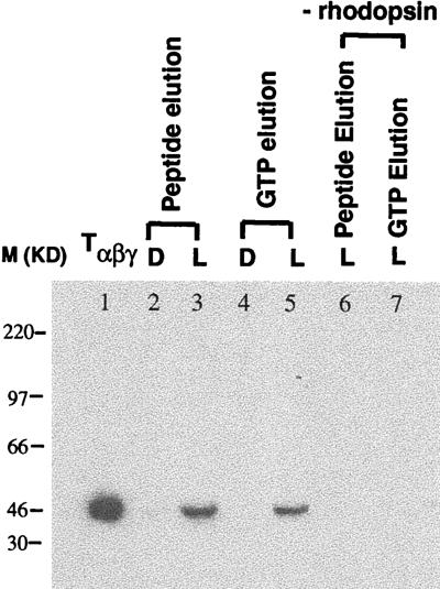
Figure 5.
Light-dependent binding of GDP/Tαβγ to wild-type rhodopsin bound to 1D4-Sepharose. Analysis was performed by SDS/PAGE (reducing). Tαβγ was visualized by immunoblotting using Ab Mab4A against Tα. Samples were treated as described in Materials and Methods. After extensive washing of 1D4 Sepharose beads both in the dark (D) and after illumination (L), Tα and rhodopsin were eluted together from the matrix with the epitope peptide (lane 2, dark; lane 3, after illumination). Tα was released also alone from the matrix by the addition of GTP (lane 4, dark; lane 5, after illumination). Controls are: lane 1, Tαβγ; lanes 6 and 7, controls of lanes 3 and 5 without 1D4-Sepharose-bound rhodopsin.

