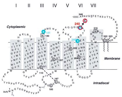
Figure 2.
A secondary structure model of rhodopsin based on the crystal structure highlighting (in red) the position of the single cysteines introduced in the cytoplasmic domain. The reactive cysteines, Cys-140 and Cys-316, in native rhodopsin were replaced by serine residues (square in black). Cysteines 167, 185, 222, and 264 are not reactive. Cys-110 and Cys-187 form a disulfide bond whereas Cys-322 and Cys-323 carry palmitoyl groups (wiggly lines).

