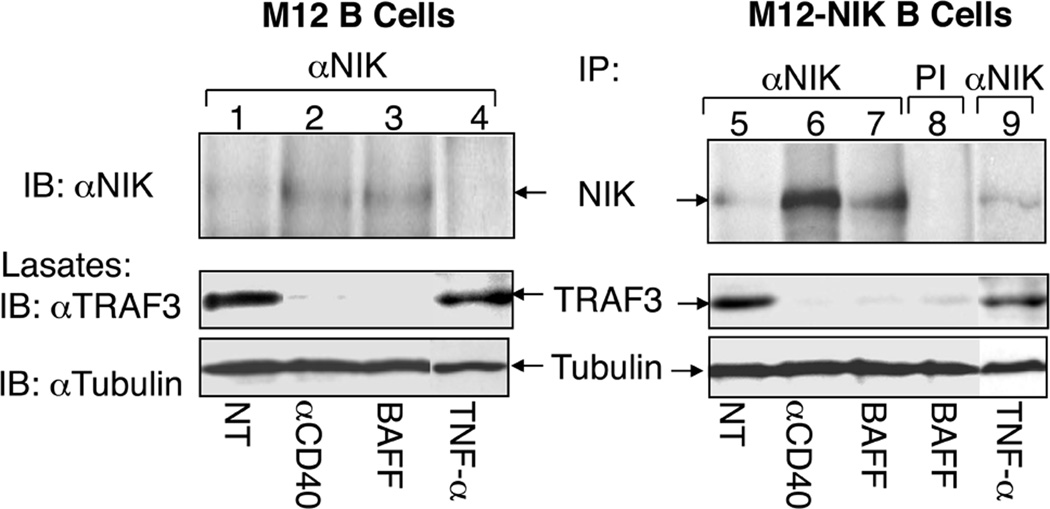
Fig. 3. Signal-induced NIK activation involves TRAF3 degradation and NIK accumulation.
M12 cells and M12-NIK cells (M12 cells stably infected with retroviruses encoding NIK) were either not treated (NT) or incubated for 2 h with anti-CD40, BAFF, or TNF-α. Whole-cell lysates were subjected to IP using anti-NIK (lanes 1–7 and 9) or a preimmune serum (lane 8) followed by detection of the enriched NIK protein by IB (top panel). The expression of TRAF3 (middle panel) and control tubulin (bottom panel) was detected by direct IB. This figure is adapted from Liao et al. (29) with permission.

