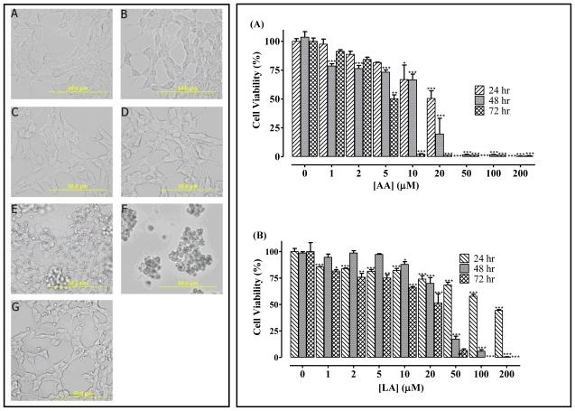
Figure 5.
left panel -AA acid induces the degeneration of human neuroblastoma SH-SY5Y cells. Human neuroblastoma cells were cultured and seeded in 24-well plates as described in the methods. They were then treated with varying concentrations of AA. Images of the cells were captured after a 24 h exposure to AA with an Olympus DP70 Camera (left panel). These show (A) untreated, (B) acetone and (C) to (F) were treated with 1, 10, 20, 50 μM AA, respectively and (G) 200 μM ARA. Right panel -AA and LA induce the degeneration of human neuroblastoma SH-SY5Y cells. Human neuroblastoma cells were cultured and seeded in 96-well plates as described in the methods. At 24, 48 and 72 h after treatment with varying concentrations of (A) AA or (B) LA as indicated, cell viability was measured by fluorescence using the resazurin reduction assay. The results are expressed as the means (± SEM, N=4) relative to the controls. *P<0.05, **P<0.01 and ***P<0.001 versus untreated control cells compared by ANOVA, followed by the Dunnett’s post-test.

