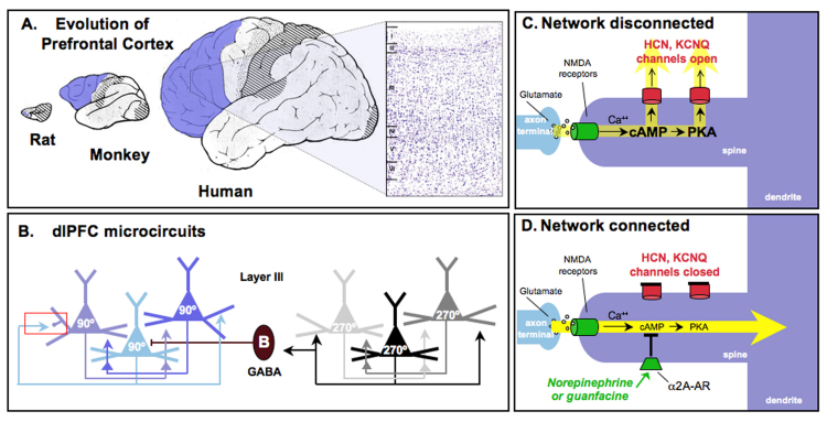
Figure 1.
Prefrontal cortical (PFC) circuits mediating higher cognitive operations. A. The PFC expands tremendously in brain evolution, comprising a very small proportion of the brain in rodents such as the rat and increasing dramatically in primates, with special prominence in the human brain. The PFC is highlighted in blue. An inset of the human dlPFC is shown at the right; this Nissl-stained section shows the six layers of dlPFC. B. The dlPFC microcircuits subserving working memory, discovered by Goldman-Rakic (1995) [1]. Pyramidal cells in deep layer III receive visuospatial information from the parietal association cortex. Pyramidal cells with similar spatial inputs excite each other through connections on dendritic spines to maintain persistent firing throughout the delay period. The spatial tuning of the neuron’s response is sharpened by lateral inhibition from parvalbumin-containing GABAergic interneurons, such as the Basket cell (B) shown in this figure. Note that chandelier cells also serve this function (not shown). The red rectangle highlights an axo-spinous synapse enlarged in C and D. These dlPFC microcircuits are the ones most afflicted in schizophrenia, where there is loss of neuropil (including loss of dendritic spines) in deep layer III [100], and reduced parvalbumin GABAergic function [101]. C. A working model of the cAMP-potassium channel signaling mechanisms in spines that dynamically weaken synaptic efficacy and gate out network inputs to the neuron. cAMP directly opens HCN channels, while cAMP activation of PKA signaling increases the open state of KCNQ channels. cAMP generated by calcium build up, e.g., feedback fatigue via NMDA or mGluR1/5, or actively generated by stress exposure, e.g., via D1 or β1 receptor stimulation. D. NE or guanfacine stimulation of α2A receptors on spines inhibits cAMP production and closes HCN and KCNQ channels, strengthening network connectivity, increasing neuronal PFC firing, and thus improving PFC regulation of behavior, thought and emotion. See Figure 5 for data supporting the model. C and D artistically adapted from Arnsten et al., 2010 [71].

