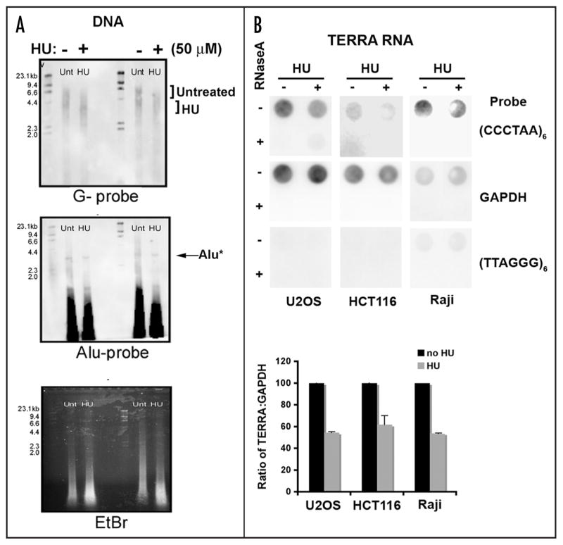
Figure 2.
Telomere dysfunction in cells treated with low dose hydroxyurea (HU). (A) Raji cells were treated with low dose HU (50 μM) for 6 days and genomic DNA was extracted and digested with Alu/MboI. A Southern hybridization with a DIG-labeled (TTAGGG)6 probe was used to assess telomere length. An Alu probe and ethidium bromide stained were used as loading controls. A DNA ladder is located on the left hand side of each gel and the numbers correspond to kilobases of DNA. (B) U20S, HCT116, and Raji cells were treated with low dose HU and total RNA was extracted. Samples were treated with (+) or without (−) RNAse. Dot-blots of RNA samples were used to assess the TERRA levels in treated cells using a DIG-labeled (CCCTAA)6 probe. Control probes for GAPDH or TERRA-complementary strand RNA (TTAGGG)6 probe were used as specificity controls.

