Figure 3.
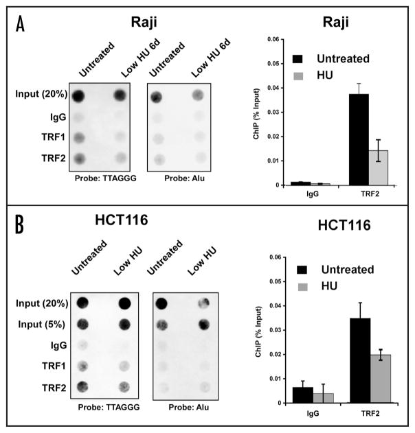
Dissociation of telomere repeat factors from telomeres after low HU treatment. (A) Raji cells were treated with low HU and then assayed by ChIP for TRF1, TRF2, and control IgG binding to telomeres. ChIP DNA was visualized by dot blot and probing with a (TTAGGG)6 (left) or Alu (right) probe. Bar graph is a summary of three independent ChIP assays as shown (A), quantified by PhosphorImager analysis and presented bound DNA relative to input DNA. (B) HCT116 cells were assayed, as in (A), by ChIP assay for TRF1 and TRF2 binding to telomere repeats (left) or Alu repeats (right). Bar graph shows quantification of three independent ChIP assays in HCT cells for TRF2 and IgG, and presented as percentage bound relative to input.

