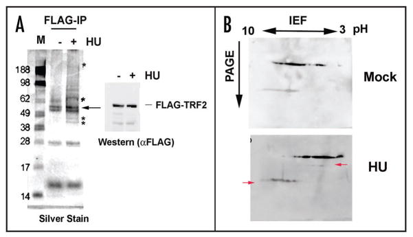
Figure 5.
Low HU alters TRF2-associated proteins and the isoelectric point of TRF2. (A) FLAG-TRF2 eluted protein was electrophoresed onto a 4–12% NuPage gel and silver stained. * represent unique bands and the arrow indicates the position of the TRF2 protein. A western blot of the input material is shown to the right of the silver stained gel. (B) FLAG-TRF2 eluted protein was used in 2D protein gel electrophoresis to determine a difference in isoelectric point of the protein after treatment. The protein was first run on pH 3–10 (non-linear) strips and then subjected to second-dimension protein electrophoresis according to molecular weight. The protein was transferred to a nitrocellulose membrane and used in western blotting. Arrows indicate changes in TRF2 mobility after treatment with HU.

