Figure 1.

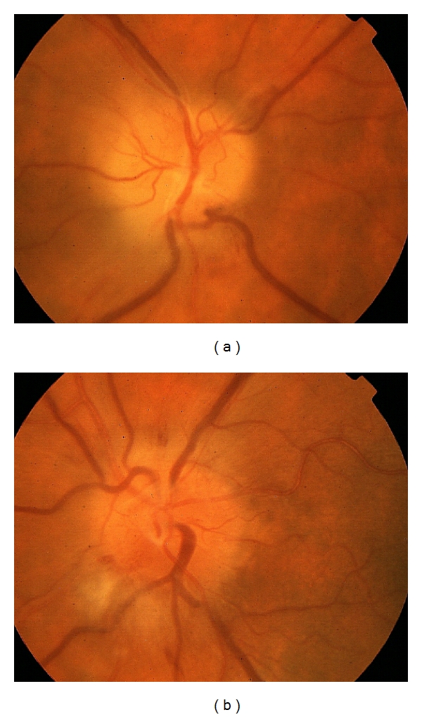
Color fundus photo of OD (a) and OS (b) with optic disc edema and peripapillary nerve fiber layer hemorrhages (a, b) and peripapillary cotton wool spots (b).

Color fundus photo of OD (a) and OS (b) with optic disc edema and peripapillary nerve fiber layer hemorrhages (a, b) and peripapillary cotton wool spots (b).