
Figure 4.
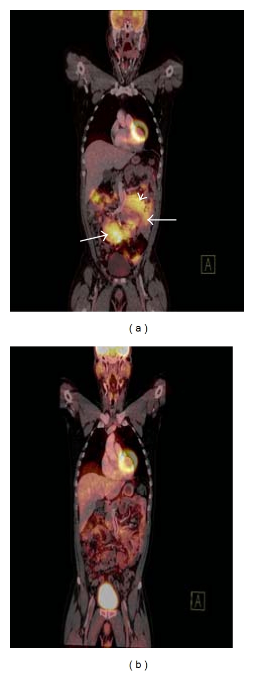
Coronal fused FDG-PET/CT image (a) demonstrating increased radiotracer uptake in multiple intra-abdominal lymph node masses in a patient with DLBCL (white arrows). Note how a large soft tissue mass displaces the small bowel and the mesenteric vessels (white arrowhead). Coronal fused FDG-PET/CT image (b) in the same patient demonstrating complete resolution of the previously described soft tissue masses. The observed residual FDG avidity is within bowel and is normal. This FDG-PET/CT confirms complete response to treatment.