
Figure 1.
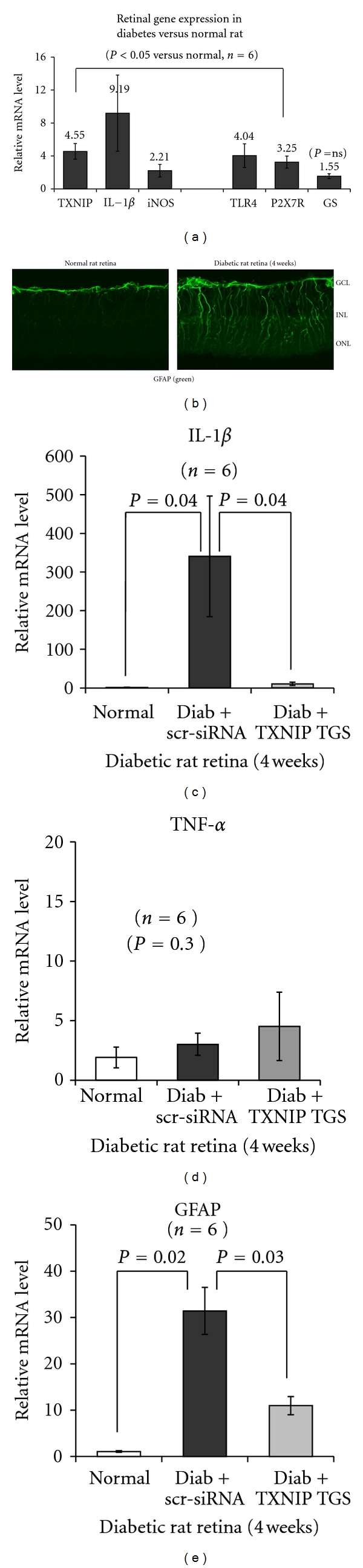
Diabetes induces TXNIP and pro-inflammatory gene expression and glia reactivity in the rat retina. (a) Messenger mRNA levels for TXNIP (4.55-fold ± 0.96) and pro-inflammatory IL-1β (9.19 ± 4.62), iNOS (2.22 ± 0.76), and pattern recognition receptors TLR4 (4.04 ± 1.43) and P2X7R (3.26 ± 0.73) are increased significantly (P < 0.05, n = 6) in the retina of diabetic rats (4 weeks) when compared with the normal retina. GS is marginally increased (1.55 ± 0.29) but not significant (P = 0.3). (b) GFAP staining, a marker of gliosis, is also increased radially throughout the neuroretina in the diabetic rat versus the normal retina, suggesting Muller glia activation. GCL: ganglion cell layer; INL: inner nuclear layer; and ONL: outer nuclear layer. (c–e) TXNIP knockdown by siRNA targeted to the promoter (RNAi TGS, [9]) reduces IL-1β and GFAP mRNA levels in the diabetic retina as compared to the scr-siRNA-treated diabetic rat retina. Under these conditions (4 weeks of diabetes induction in rats), we did not see an increase in retinal TNF-α mRNA level.