
FIGURE 3.
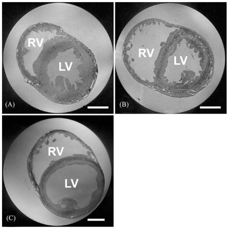
MR image planes obtained from contractured (A), slack (B), and volume-loaded (C) hearts (scale bar: 5 mm). In (A) and (B), voxel size is 26.5 × 26.5 × 24.5 μm, and in (C) 32 × 32 × 44 μm (a larger bore NMR tube was required for the volume-loaded heart). RV: right ventricle; LV: left ventricle. A full movie of the data in Figure 3B, progressing through transversal MRI sections from aorta to apex, can be downloaded from: http://mef.physiol.ox.ac.uk/MRI/Aorta_to_apex.avi.