FIG. 1.
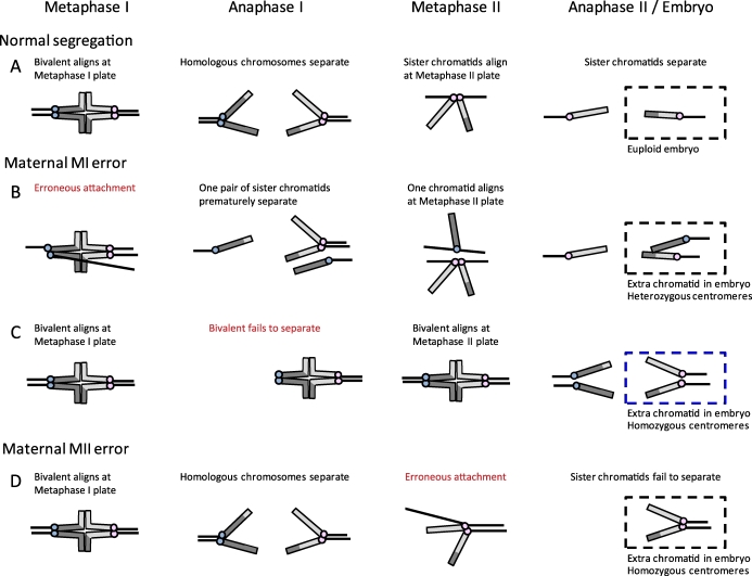
Analysis of centromeres to determine the origin of maternal errors in trisomies. Schematic of chromosome segregation in MI and MII. Chromosomes are depicted as telocentric (as in mice) for simplicity, and centromeres of sister chromatids are colored alike (e.g., blue or pink). A) Normal segregation at both MI and MII, with a euploid embryo. B) Example of an error at MI, where a pair of sister kinetochores erroneously biorients at metaphase I and prematurely separates at anaphase I; the resulting chromatids in the embryo have heterozygous centromeres. Note that there are several possible segregation errors that can occur at MI (e.g., because of failure of recombination or loss of cohesion), of which one example is shown, but heterozygous centromeres in the embryo indicate that an MI error occurred. C) Example of an error at MI, where a bivalent fails to separate at anaphase I and instead separates at anaphase II. Here the centromeres in the embryo are homozygous and would be problematically classified as a MII error, even though the initial error occurred in MI. D) Example of an MII error, where sister chromatids fail to separate at anaphase II; the resulting chromatids in the embryo are homozygous.

