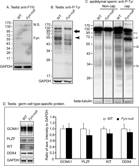
FIG. 1.
Western blot analysis of Fyn-null testis and spermatozoa is shown. Testes were homogenized in RIPA buffer, and sperm were lysed in Laemmli sample buffer. After centrifugation, soluble protein was analyzed by Western blotting as described in Materials and Methods. A) The expression levels of FYN kinase in the testis of WT and Fyn-null mice were detected with an anti-FYN antibody (NS, nonspecific band). B) Patterns of P-Tyr-containing proteins in WT and Fyn-null testes were detected with anti-P-Tyr antibody. Anti-GAPDH was used as a loading control. An arrow (close to 95 kDa) and arrowhead (approximately 55 kDa) indicate P-Tyr-containing proteins in the WT but not in the Fyn-null sample. C) Patterns of P-Tyr-containing proteins in WT and Fyn-null sperm were detected in noncapacitating (Non-cap) and capacitating (cap) sperm by using an anti-P-Tyr antibody. P-Tyr-containing proteins at ∼95 kDa (i), ∼72 kDa (ii), ∼50 kDa (iii), or ∼30 kDa (iv) exhibited reduced antibody binding in Fyn-null capacitating sperm relative to those of WT. D) Analysis of the expression levels of different germ cell type-specific proteins in the testis is shown. Testis proteins from WT and Fyn-null males were analyzed by Western blotting with antibodies to GCNA1 (representing germ cells, except for elongating spermatids), PLZF (spermatogonial stem cells or undifferentiated spermatogonia), KIT and DDX4 (differentiating/differentiated germ cells), and GAPDH (a loading control). The bar graph represents the ratios between average intensity of identified protein band and that of GAPDH and WT control. Values represent means ± SD for six testes in a Fyn-null or WT group. Comparison by t-test indicated that individual expression levels of the four analyzed proteins of Fyn-null mice were not significantly different from those of WT mice.

