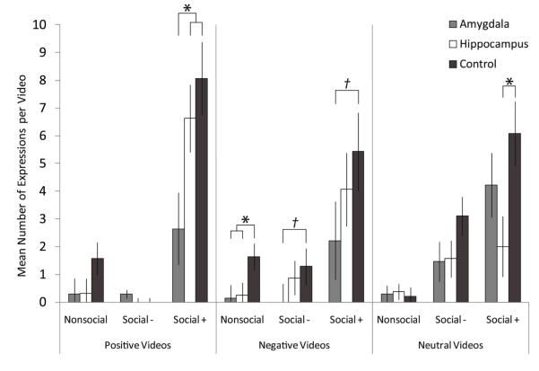
Figure 2.
Expressions Directed Towards Video
Data columns depict the mean number of expressions per video type; error bars represent standard errors of the estimated marginal means. Amygdala: amygdala-lesioned animals; Hippocampus: hippocampus-lesioned animals; Control: control animals. Baseline: baseline trials with screen saver video. Affective Content Categories—Positive: videos with positive affective content; Negative: videos with negative affective content. Neutral: videos with neutral affective content. Social Content Categories—Nonsocial: Nonsocial videos. Social−: Social non-engaging videos. Social+: Social engaging videos. * indicates significant between group differences based on post-hoc LSD comparisons or t-tests. † indicates marginally significant (trend) between group differences based on post-hoc LSD comparisons or t-tests.
Amygdala-lesioned animals were significantly less responsive to positive social engaging videos than hippocampus-lesioned and control animals, F(2, 22)= 4.68, p<.023, np2=.330 (amygdala-lesioned < hippocampus-lesioned, control, p<.05). Amygdala- and hippocampus-lesioned were marginally less responsive than controls to negative nonsocial videos, F(2, 22)= 3.01, p<.07, np2=.245 (amygdala-lesioned, hippocampus-lesioned < control, p<.05). Post-hoc tests following a marginally significant effect of lesion condition on responsivity neutral social engaging videos F(2, 22)= 3.279, p<.06, np2=.257, revealed that hippocampus-lesioned animals were significantly less responsive than controls (p<.05). Amygdala-lesions animals were marginally less responsive than controls to negative social nonengaging videos, t(6)=2.12, p<.08, and negative social engaging videos t(12)=2.12, p<.06.

