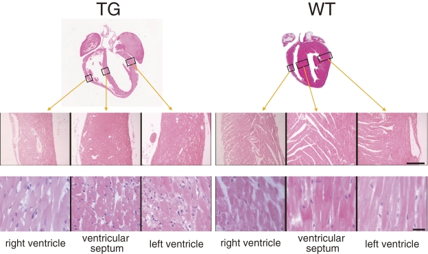
Figure 4.
Histopathological observations of hearts in 6-month-old transgenic (TG) and wild-type (WT) mice. Hematoxylin and eosin-stained tissue sections from three cavity walls, the positions of which are indicated by rectangles in the upper images and are shown in low (middle images, bar = 100 µm) and high (lower images, bar = 10 µm) magnifications. Slight fibrosis was observed sporadically in TG hearts, but no apparent cell infiltration was found in either TG or WT hearts.

