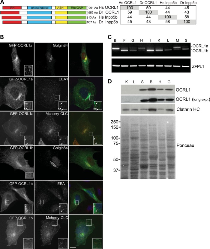
Figure 1.
OCRL1 domain organization, subcellular localization, and tissue-specific splicing and expression are conserved in zebrafish. (A) Schematic view of human and zebrafish OCRL1 and Inpp5 showing the predicted PH, 5-phosphatase, ASH and RhoGAP-like domains. The table indicates amino acid identity between the proteins. (B) Immunofluorescence microscopy of zebrafish AB9 or PAC2 fibroblast cells expressing GFP-tagged OCRL1 isoform a or b (green) with or without mCherry-tagged clathrin light chain (CLC) and labelled with antibodies to Golgin84 or EEA1 (red). Arrows indicate colocalization of OCRL1a with EEA1 or mCherry-clathrin light chain. Scale bar, 10 µm. (C) RT–PCR of adult zebrafish tissues using primers against OCRL1 isoform a and b, or the control ZFPL1. B, brain; F, fin; G, gill; H, heart; I, intestine; K, kidney; L, liver; M, muscle; S, skin. (D) Western blot of zebrafish adult tissues using the indicated antibodies. K, kidney; L, liver; S, skin; B, brain; H, heart; G, gill. Ponceau staining of the membrane shows similar loading per lane.

