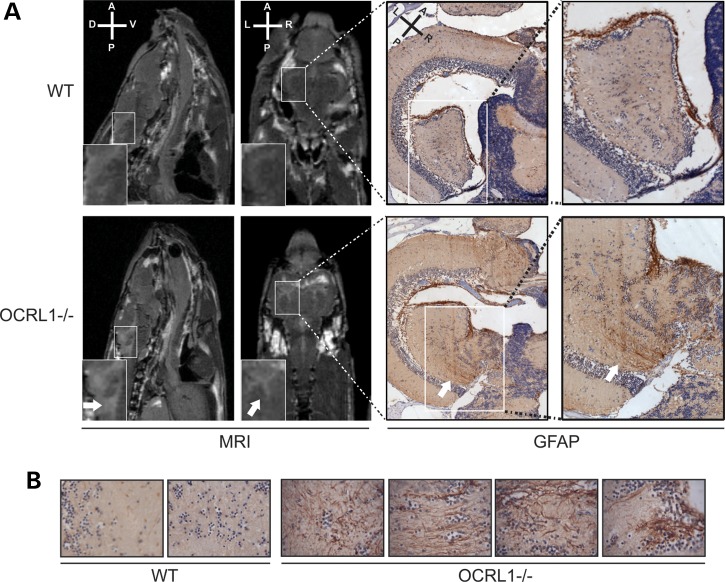
Figure 5.
White matter lesions in OCRL1 mutant zebrafish. (A) Transverse relaxation (T2)-weighted MRI images of the brains of WT and OCRL1−/− adult zebrafish. Sagittal images indicate the presence of a white matter anomaly (white arrow in insert) in OCRL−/−, which was not seen in WT. Coronal images show the presence of lesions adjacent to the ventricles (white arrow in insert). Immunohistochemistry was carried out on the same samples used for MRI. A higher number of GFAP-positive astrocytes are present in the periventricular lesion suggesting increased gliosis. (B) Examples of GFAP staining of WT and OCRL1−/− brain regions. Note the increased numbers of astrocytes concentrated in regions that correspond to periventricular lesions of OCRL1−/− embryos.

