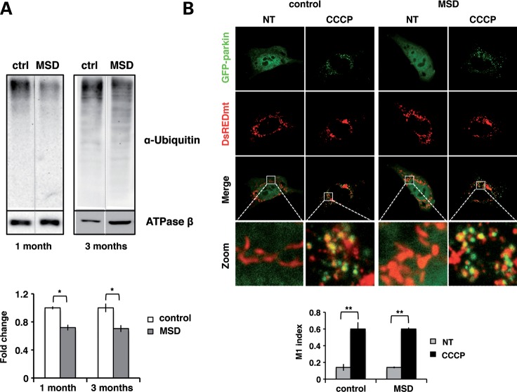
Figure 4.
Incomplete ubiquitination is not due to impaired parkin translocation to mitochondria. (A) Anti-ubiquitin immunoblots of liver mitochondrial extracts obtained from MSD (n = 4) and control (n = 4) mice at 1 month and 3 months. ATPase β was used as mitochondrial loading control and results were expressed as the fold changes of the ubiquitin/ATPase β ratio; *P < 0.05. (B) Confocal images (63×) of untreated (NT) and CCCP-treated (20 μm, 20 h) MSD and control MEFs co-expressing GFP-parkin (green) and DsREDmito (red) plasmids. The zoomed region is indicated with a white square. Co-localization is expressed as the M1 (Mander's) index. ANOVA P-value = 0.158, **P < 0.01.

