
Figure 1.
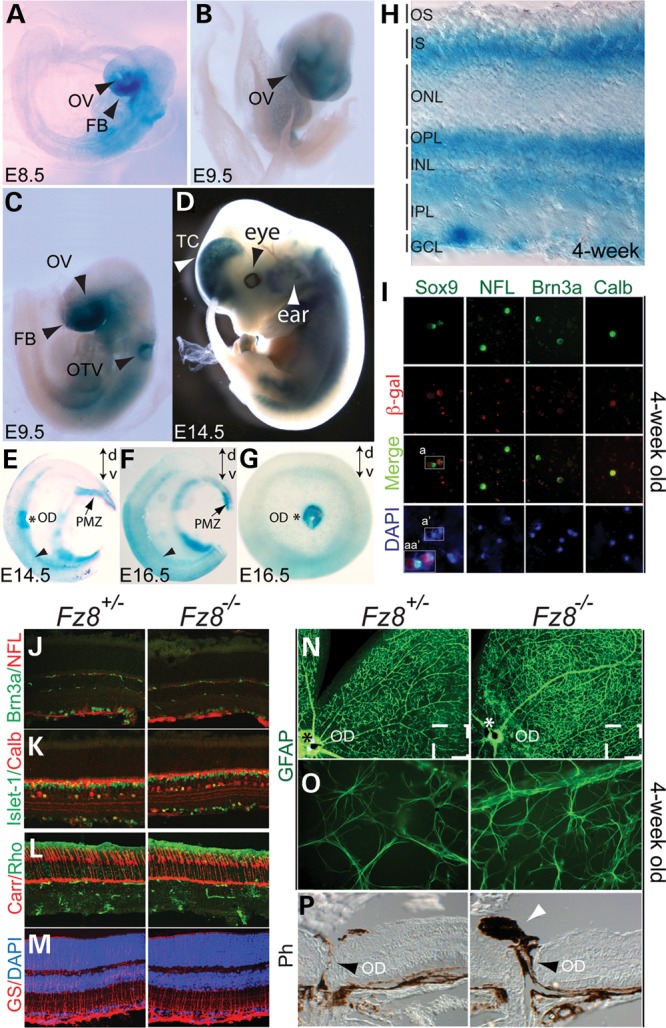
Fz8 retinal expression during mouse development and phenotypic analysis of Fz8 mutant retina. (A–H) Fz8 expression was visualized by a β-gal reporter from its knock-in endogenous locus. (A–C) Fz8 was expressed in the areas of embryonic forebrain (FB), optic vesicle (OV) and otic vesicle (OTV) at early stages of embryonic day 8.5 (E8.5) and E9.5. (D) Fz8 expression was continuously seen in forebrain ear and eye regions at E14.5. (E and F) Bisected X-gal stained whole-mount retinas at E14.5 (E) and E16.5 (F) showing Fz8 enhanced expression in optic disc (OD, asterisk), PMZ (arrows) and ventral retina (arrowhead). Dorsal–ventral orientation (d-v) is indicated by up-down arrows in each panel. (G) A top view of an X-gal-stained E16.5 retina showing a preferential Fz8 expression in optic disc and ventral retina. (H) A 4-week-old X-gal-stained retinal section shows that Fz8 was expressed through all retinal layers. OS, outer segment; IS, inner segment; ONL, outer nuclear layer; OPL, outer plexiform layer; INL, inner nuclear layer; OPL, outer plexiform layer; IPL, inner plexiform layer; GCL, ganglion cell layer. (I) Co-immunostaining using anti-β-gal antibody (red) and other retinal cell markers (green) shows that Fz8 is expressed in Müller cell, ganglion cell (RGC) and Calbidin-positive cells (likely amacrine cells). Merged images show co-labeling of β-gal with Sox9 for Müller cell, NFL and Brn3a for ganglion cell, and Calbindin (Calb) positive cells. DAPI staining (blue) shows number of cells in the pictured field. Boxed areas indicate that same cells are being labeled by Sox9 and β-gal. a, double-labeled cells with Sox9 and β-gal; a′, DAPI-stained cell nuclei; and aa″, zooming in on merged channels of Sox9, β-gal and DAPI in a and a′ areas. (J–M), normal development of retinal cell types in Fz8 mutant retinas: no significant differences in immunostained Fz8 heterozygous and mutant retinas for Brn3a- and NFL-positive RGCs (J, green and red are for RGCs and RGC axons, respectively), Islet-1-positive RGCs, ACs and bipolar cells (K, green), calbindin (Calb)-positive ACs and HCs (K, red), rhodopsin-labeled rods (L, green), cone arrestin (Carr)-labeled cones (L, red) and glutamate synthetase (GS)-labeled Müller glia. Non-specific staining (green signals in IPL and OPL) in (J) and (L) is from anti-mouse Ig secondary antibody. (N–P) Increased GFAP staining of astrocytes dendrites in the mutant retina, and mild optic disc defects. (N) Low magnification showing the GFAP stained whole-mount retinas showing enhanced GFAP staining. (O) Zoom-in areas roughly correspond to the boxed areas in (N), showing individually stained astrocytes dendrites. (P) A frequent observation (6/11 mutant mice) of optic disc defects associated with the enhanced GFAP staining, featured by PFV (white arrowhead). The optic disc (OD, black arrowhead) does not seem to close properly in the mutant mice.