
Figure 5.
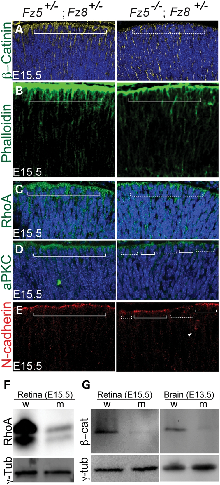
Retinal apical junction is perturbed in developing the Fz5−/−;Fz8+/− mutant retina. (A) Mislocalization of β-catenin in the mutant retina. β-Catenin localization is enriched on the apical retina (left panel, solid bracket), but became more homogenous in the mutant retina. The DAPI-stained apical nuclei in the WT are well topped by β-catenin-enriched apical cytoplasm, while they are seemingly more naked in the mutant (dashed bracket). (B) In the WT E15.5 retina, phalloidin-stained F-actins are enriched on the apical retinal surface (bracket, left panel), while in the mutant retina less staining is seen (right panel). (C) Reduction and discontinuity of RhoA enrichment on apical surface of the Fz5−/−;Fz8+/− mutant retina (dashed bracket). (D) Discontinuity of aPKC on the apical junction of the mutant retina (compare regions above solid and dashed brackets in the mutant retina). (E) Discontinuity of N-cadherin staining at the mutant retinal apical junctions. Ectopic N-cadherin staining is also sometimes seen in the subapical area in the mutant retina (arrow in right panel), but not in the WT. (F and G) Western blots using whole retina and brain tissue extracts of E15.5 embryos showing significant down-regulation of RhoA and β-catenin at protein level. w, wild-type; m, mutant. Retinal sections were cut through the central–ventral area horizontally, where the mutant retinal phenotype is readily obvious under the microscope. Each panel represents five projected images at 1 μm step. All mutant retinal sections are shown in right panels.