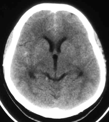
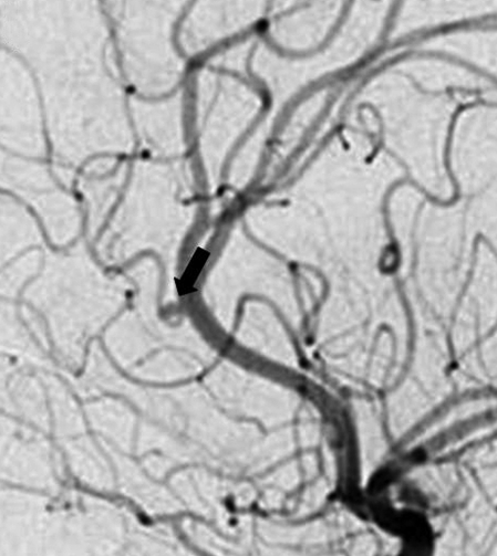
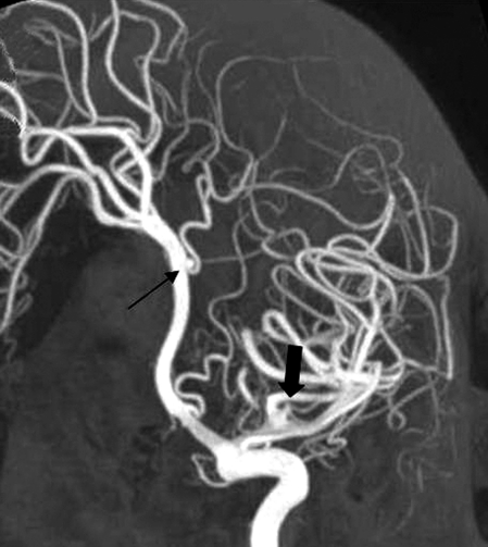
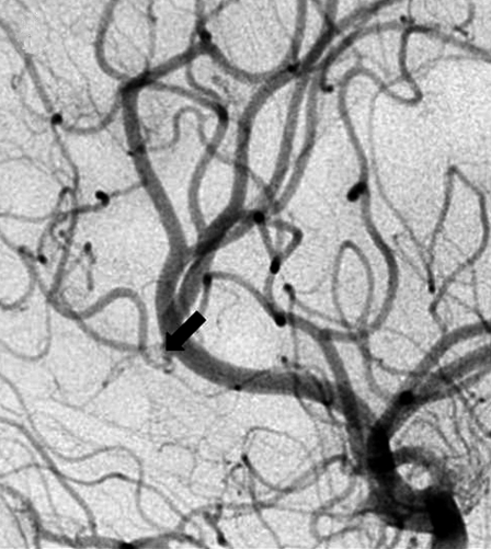
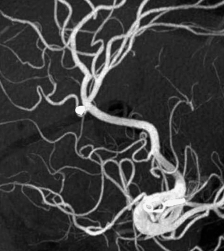
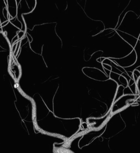
Figure 1.
A) Noncontrast head CT at admission. Note subarachnoid hemorrhage in the interhemispheric fissure and the left sylvian fissure. Left sylvian fissure is not visible at this section due to slight asymmetry of the head position. B,C) Angiography images. B) Close-up lateral view of the left internal carotid artery injection, small distal azygos anterior cerebral artery (AACA) aneurysm with a 1.5 mm diameter on the long axis is shown by the arrow. Note also the branch arising close to its neck. C) 3D angiography sagittal oblique MIP view, thin arrow indicating the distal AACA aneurysm, thick arrrow shows the MCA aneurysm. D-F) Control angiography three months after the discharge. D) Close-up lateral view, left ICA injection arrow shows the aneurysm site. E) Oblique sagittal MIP view of the 3D angiography, coils in the AACA and clip on the MCA aneurysms are seen. F) Volume rendered image, the position of the clip is seen with no residual filling in the MCA aneurysm.

