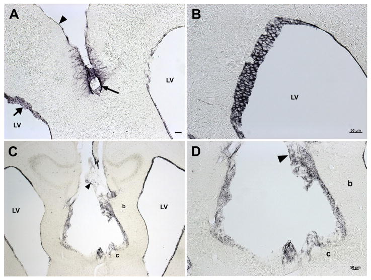
Figure 5.
3H1 wild-type and affected mouse brains immunostained for vimentin. (A) Coronal section of 3H1 wild-type positive for immunostaining staining vimentin in meninges (arrowhead), tanycytes (arrow) and ependymal cells (broad arrow) lining the lateral ventricles (LV). (B) Higher magnification of ependymal cells of the lateral ventricle from the 3H1 wild-type mouse shown in (A). (C) Affected 3H1 tuft immunopositive in adipose tissue of the interhemispheric lipoma (arrowhead), ependymal cells, but negative in the fibrous capsule (c) and brain parenchyma (b). (D) Higher magnification of lipoma in (C) showing positive staining for vimentin in adipose tissue (arrowhead) but negative in adjacent capsule (c) and parenchyma (b). Bar = 50 microns.

