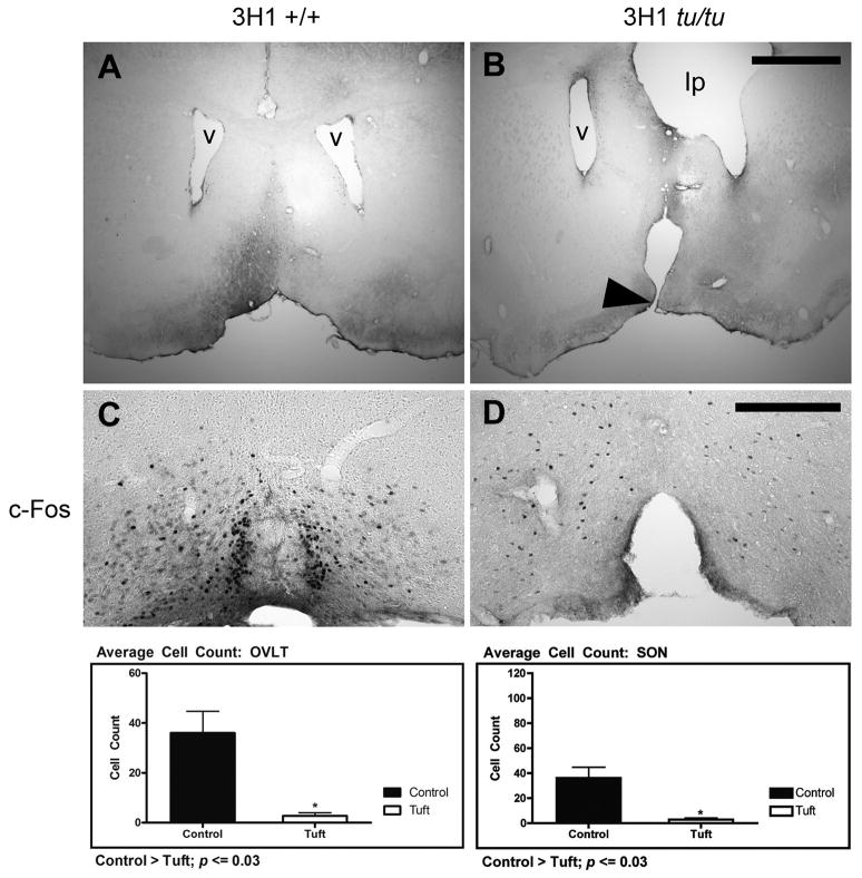
Figure 6.
Malformation of the lamina terminalis. (A) Coronal section of 3H1 wild-type at the level of the lateral ventricles with a normal lamina terminalis (B) 3H1 tuft showing discontinuity along the basal forebrain region corresponding to an abnormally formed lamina terminalis (arrowhead). Distorting effects on one of the lateral ventricles (v) by the intracranial lipoma (lp) was also revealed in one of the affected mice with a lipoma. Representative c-Fos immunostaining in (C) 3H1 wild-type and in (D) 3H1 tuft mouse serial sections from the mice in (A) and (B) respectively. (Bottom) Bar graphs illustrating the difference in cell numbers expressing c-Fos within the OVLT and SON averaged from a sample number of five. Bar = 1.0 mm for A and B. Bar = 200 microns for C and D.

