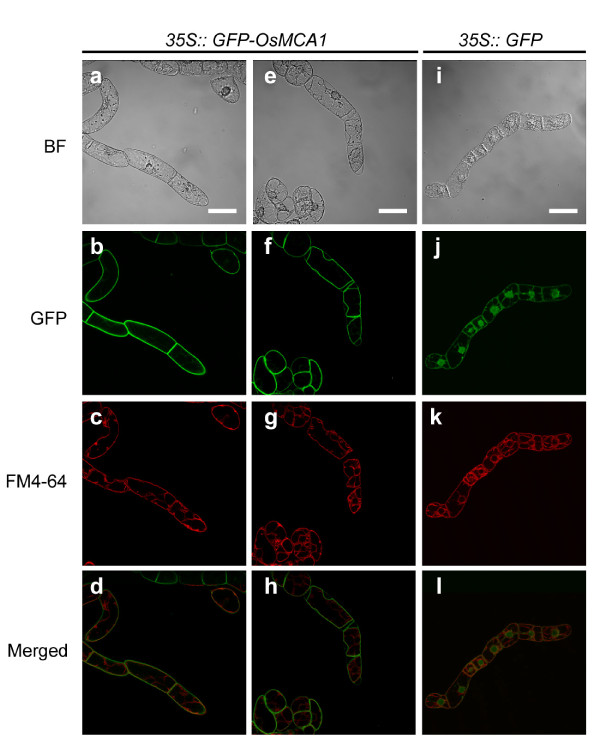
Figure 2.
Intracellular localization of the OsMCA1 protein. Confocal fluorescence images (b-d, f-h, j-l) and differential interference contrast (DIC) images (a, e, i) of tobacco BY-2 cells expressing GFP-OsMCA1 (a-h) or GFP (i-l) stained with FM4-64 (4.25 μM) for 3 h. Fluorescence of GFP (b, f, j) and FM4-64 (c, g, k). (e-h) Plasmolyzed cells. Scale bar: 20 μm.

