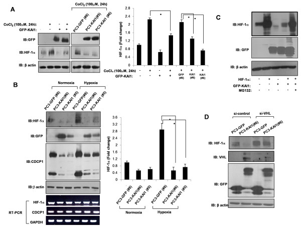
Figure 4.
KAI1 blocks HIF-1α induction. (A) After transfection of KAI1, cells were treated with 100 μM CoCl2 for 24 hours. PC3 vector control (PC3-GFP #8) and KAI1 transfectants (PC3-KAI1 #5 and PC3-KAI1 #6) were also treated with 100 μM of CoCl2 for 24 hours. Cells were harvested and immunoblotted for detection of HIF-1α and GFP-KAI1. Densitometric analysis of HIF-1α was shown after normalization to beta-actin. (B) PC3 vector control (PC3-GFP #8) and KAI1 transfectants (PC3-KAI1 #5 and PC3-KAI1 #6) were exposed to hypoxia for 24 hours. The levels of HIF-1α, CDCP1, and GFP-KAI1 proteins were assessed by immunoblotting. Densitometric analysis of HIF-1α was shown after normalization to beta-actin. HIF-1α and CDCP1 mRNA levels were measured by RT-PCR. (C) PC3 cells were cotransfected with KAI1 and HIF-1α, then treated with the proteasome inhibitor, MG132. HIF-1α levels were assessed by immunoblotting. (D) Stable KAI1-expressing (PC3-KAI1 #6) and vector control (PC3-GFP #8) PC3 cell clones were treated with siRNA against CDCP1 and VHL. Forty-eight hours after transfection, HIF-1α, CDCP1, and VHL protein levels were measured by immunoblotting.

