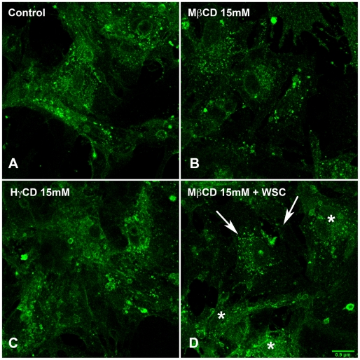
Figure 3. Treatment with MβCD leads to changes in membrane raft organization of cardiomyocytes.
Confocal images of control (A) and cardiomyocytes pre-treated with 10 mM MβCD (B) or HγCD (C). Cells were washed, fixed and then labeled with CTXb-Alexa 488, which recognizes GM1, a raft marker. In comparison to control cells, which show a homogenous strong labeling for GM1, cholesterol-depleted cardiomyocytes reveals a more discrete labeling. Cells treated with HγCD show GM1 labeling similar to control cells whereas cholesterol-replenished cells (D) exhibit both patterns of cholesterol-depleted (arrows) as well as control (asterisks) GM1 labeling. Scale bar: 0.9 µm.

