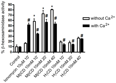
Figure 4. MβCD but not HγCD cell incubation leads to lysosomal exocytosis in cardiomyocytes.
Cardiomyocytes were exposed to either 10 mM MβCD or HγCD for 10, 20 or 40 minutes at 37°C, in the absence (white bars) or presence (black bars) of calcium. Both extracellular media and lysates were collected and exposed to 4-methylumbelliferyl-N-acetyl-B-D-glucosaminide, the fluorescent substrate of beta-hexosaminidase, an enzyme resident within lysosomes. Results are shown as ratio between β-hexosaminidase activity in extracellular media and total β-hexosaminidase activity (extracellular media over extracellular media plus β- hexosaminidase cell lysate hexosaminidase activity). Cells treated with 10 µM Ionomycin (Calbiochem) for 10 minutes were used as lysosomal exocytosis positive control. Data are shown as mean of triplicates ±SD. Asteriks indicate statistically significant differences (p < 0.05,Student's t test) between control and treated cells.

