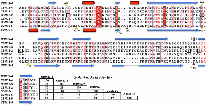
Figure 5. Amino acid sequence comparison of the CBM32 modules from CpGH89.
The secondary structure is shown above (CBM32-4) and below (CBM32-5) with arrows representing β-strands and cylinders α-helices. The purple and yellow triangles above and below the sequences indicate the aromatic and hydrogen bonding residues, respectively, that are involved in carbohydrate binding by CBM32-4 (top) and CBM32-5 (bottom). Numbers with the triangles indicate the residue number. Residues in CBM32-2 that are highlighted by boxes are those present in the putative binding site of this module.

