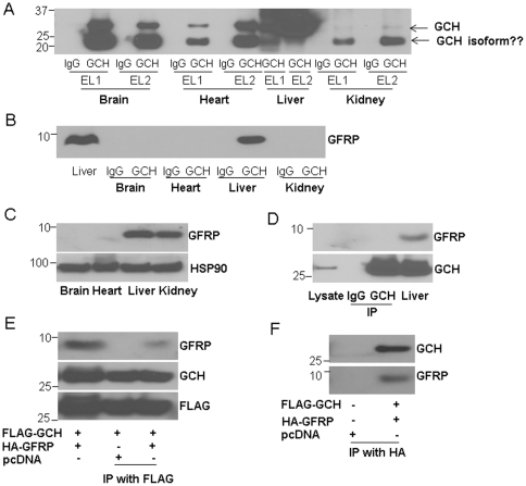
Figure 4. The interaction of GCH1 with GFRP in different organs.
GCH1 and its interacting proteins were purified from brain, heart, liver and kidney, and analyzed by western blot against GCH1 antibody (A) and GFRP antibody (B). EL1 is the first eluate, EL2 is the second eluate. Eluates from IgG conjugated column were used as controls. Protein lysates from different organs were immunoblotted with GFRP antibody and HSP90. HSP90 was used as a loading control (C). HEK cells stably over-expressing GCH1 (GCH1-HEK) were immunoprecipitated with IgG or GCH1 antibody and immunoblotted against GFRP and GCH1. Straight cell lysate of GCH1-HEK cells (Lysate) was also loaded for comparison (the first lane). Liver homogenate was used as a positive control (D). HEK cells were transiently transfected with Flag-GCH1, HA-GFRP or pcDNA, and immunoprecipitated with Flag tag (E) or HA tag and immunoblotted with GCH1 and GFRP antibodies. In (E), cell lysates (the first lane) from HEK cells transfected with FLAG-GCH1 and HA-GFRP were used as positive controls for GFRP and GCH1 expression.

