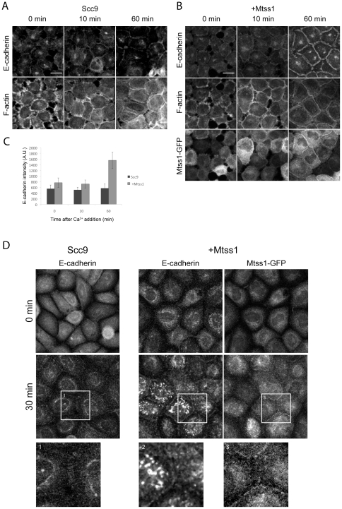
Figure 2. Mtss1 enhances de novo cell-cell junction formation.
(A, B) Scc9 cells +/− Mtss1-GFP were treated with 2 mM EGTA for 20 min, to disrupt junctions. Reassembly was stimulated by 2 mM Ca2+ and cells were labeled for E-cadherin and F-actin. E-cadherin and F-actin were visualized following triton extraction and fixation to preserve the triton-insoluble junctional cytoskeleton. (A) Cell-cell junction formation in Scc9 cells and (B) Mtss1-GFP expressing Scc9 cells (C) Mean intensity of E-cadherin fluorescence at cell-cell junctions in Scc9 cells ± Mtss1 (mean ± S.D) from 3 independent experiments where n = 20 junctions. (D) Scc9 cells ± Mtss1-GFP cultured in low-Ca2+ KSFM overnight to disassemble adherens junctions. Cells were Ca2+ treated, fixed and labeled after the indicated times. Enlarged images below show boxed regions.

