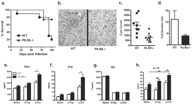
Figure 1. Characterization of the local inflammatory response during chronic i.p. infection.
WT and Pilrb −/− mice were challenged i.p. with 20 cysts T.gondii and followed over time. Survival of WT and Pilrb−/− mice through 100 days post infection (a). H&E of brain sections reveal larger inflammatory foci in WT mice compared to Pilrb−/−, shown at 10× magnification (b). Total number of cysts present in the CNS of mice 60–90 days post infection (c). Actual numbers of BMNCs isolated from the CNS of mice (d). Recall assays using BMNCs isolated from WT and Pilrb−/− mice and cultured for 72 hrs in the presence of Media alone, STAg, or αCD3. Levels of protein are shown as detected by ELISA for TNFα (e), IFNγ (f), NO (g), and IL-10 (h). For panel a, n = 5–15 mice/group for each of 3 experiments performed. For panel c, pooled data from 2 experiments are shown, p<0.004. For panel d, one representative experiment of 2 is shown, p<0.05. For panels e,f, and h, pooled data from 2–3 experiments are shown, *p<0.005; **p<0.002.

