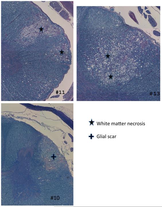
Figure 4.
Examples of individual retreated pigs. The top two images represent responders after 20Gy (#11 & 13), each showing focal white matter necrosis in the dorso-lateral white matter on the reirradiated side. The lower picture shows a focal area of glial scarring in an 18Gy non-responder (#10) one year after retreatment.

