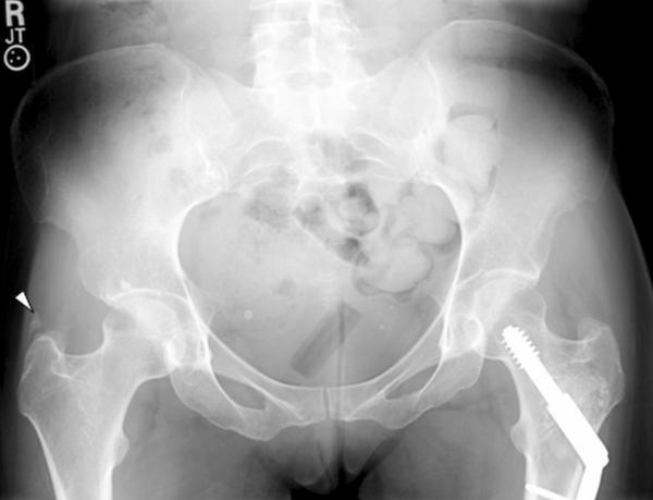
Figure 3.
Pelvic radiographs. Anterior-posterior radiograph of the pelvis.
Note normal joint spaces of the hip joints on both sides without obvious degeneration. On the right side, adjacent to the greater trochanter there is an abnormal ossicle [arrow] suggesting tendinopathy of the gluteal muscles. The left femur demonstrates postsurgical changes with osteosynthetic treatment of the femoral neck.

