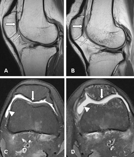
Figure 4.
MR imaging of the knee joints.
Sagittal [A, B] and transverse [C, D] images of the right [A, C] and left [B, D] knee joint, respectively. On the right side, absence of the articular cartilage [thin arrow] of the femoral joint surface [A] and marked defects and thinning of the retro-patellar cartilage [thick arrow]; whereas the cartilaginous layers [arrows] of the left knee are only mildly thinned [B]. The transverse images feature joint effusion on both sides [arrowheads] [C, D], and again distinct cartilage damage of the right retro-patellar surface [arrow] [C], and slight cartilaginous changes left-sided [arrow] [D].

