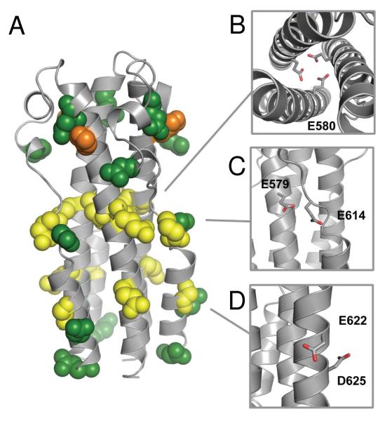
Figure 4.
(A) Structural homology model of MARV GP2 ectodomain based on the crystal structure of EBOV GP2 (ref. 11, PDB ID 2EBO). Glutamic acid (yellow), aspartic acid (green), and histidine (orange) residues are shown in spacefill. (B-C) Predicted interactions that could destabilize the six-helix bundle, shown in stick and colored by atom for clarity: (B) E580 interactions in the NHR core trimer; (C) E579-E614 interaction between opposing monomers; (D) E622-D625 i→i+3 intrahelical interaction.

