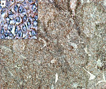
Figure 2.

Immunoperoxidase for CD99 (10×) showed the tumor composed of small round cells with round nuclei and scant cytoplasm arranged in cohesive lobules. There are spindle cellular elements with diffuse CD99 cytoplasmic staining. Inset a (40×) shows Homer-Wright type rosettes positive for CD99.
