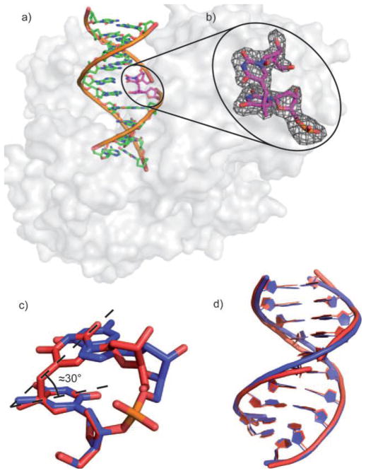
Figure 1.
a) Crystal structure of the dinucleoside SP-containing duplex in complex with B. st. DNA polymerase. b) Zoom-in-view of the dinucleoside SP which contains an R chiral center at the C5 carbon. c) Overlay of the SP lesion (red) and of an undamaged TpT segment (blue). d) Overlay of the undamaged DNA and the SP- containing DNA bound to the polymerase. (The figure is copied from reference 66 with permission from John Wiley & Sons)

