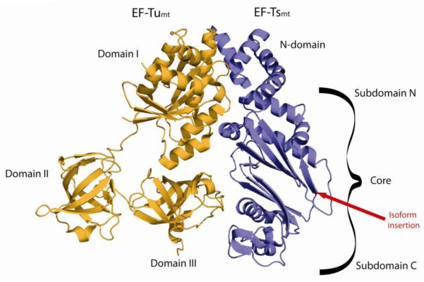
Figure 9.
Crystal structure of the bovine EF-Tumt:EF-Tsmt complex. In the 3-D structure of the bovine EF-Tumt:EF-Tsmt complex (PDB coordinates 1XB2) [101], EF-Tumt is shown in orange and EF-Tsmt is in blue. The domains of each protein are labeled and the position of the insertion present in one isoform of EF-Tsmt is indicated by a red line.

