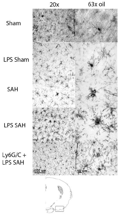
Fig. 2.

Immunohistochemistry of microglia 1 day after SAH at two magnifications (antibody, Iba1). Sham animals show rest appearing microglia with multiple long fine cell projections. LPS Sham mice show slightly more retracted processes (a sign of activation). SAH animals show ameboid morphology where the process retraction is severe, and the cells take on the appearance of macrophages. LPS SAH animals show even more densely packed projections and rounding of the cell bodies. Finally, in animals that are given a myeloid cell-depleting antibody again Ly6G/C, the microglia animals again exhibit a resting phenotype seen in the Sham animals. The diagram at the bottom shows the area of the brain imaged
